3D CBCT Imaging
Dental Technology
3S CBCT is precisely tailored to the everyday routines of private practices: it can capture the patient’s whole jaw in a single span. The field of view is large enough to avoid the stitching of several 3D x-ray images and thus multiple exposure to radiation. Yet it is also small enough to be a time-saver in diagnosis.
3D CBCT Imaging at Deer Ridge Dental
CBCT is a type of computed tomography (CT) scan that uses a cone-shaped X-ray beam to produce detailed 3D images of the oral and maxillofacial structures. Unlike traditional dental X-rays or 2D imaging, CBCT captures volumetric data (3D data), allowing the dentist to see both hard and soft tissues in much greater detail.

Highly detailed 3D Visualization
CBCT imaging offers superior detail compared to traditional 2D X-rays. It provides full 3D visualization of your teeth, bones, and soft tissues, allowing dentists to see problems that might not be visible on standard X-rays. This is especially beneficial for complex cases like impacted teeth, cysts, tumors, or bone abnormalities.
Accurate Treatment Planning
3D imaging helps dentists plan treatments with greater precision. Whether you’re getting a dental implant, undergoing orthodontic treatment, or having oral surgery, the detailed 3D images enable the dentist to map out your treatment accurately. This improves the success rate and reduces complications.
For implants, CBCT helps the dentist assess bone density, volume, and quality to determine the best placement for implants, ensuring a better fit and reduced risk of implant failure.Less Radiation
CBCT scans use less radiation than traditional CT scans, making them a safer option for patients. They typically deliver only about the same amount of radiation as a traditional panoramic X-ray or less, depending on the size of the scan area.
While all X-rays use some form of radiation, CBCT is designed to provide more detailed images with minimal exposure.
Improved Diagnosis and Detection
With 3D imaging, dentists can see problems before they become obvious, such as hidden infections, bone loss, and abscesses. It’s particularly helpful for detecting:
- Impacted teeth (like wisdom teeth)
- Hidden cavities in the roots of teeth
- Jawbone deformities or abnormalities
- Sinus issues that may affect dental treatment
- Tumors or cysts
- Nerve locations to avoid in surgeries
3D imaging als provides the ability to zoom into specific areas of interest and adjust the image contrasts makes it easier to identify and diagnose issues early.
Enhanced Implant Planning
One of the most common uses of 3D CBCT is in planning for dental implants. With 3D imaging, the dentist can assess:
- Bone density and quality in the implant area
- Precise measurements of available bone for implant placement
- The proximity to important structures (like nerves and sinuses)
- Optimal placement angles for the implant
This leads to more accurate placement of dental implants, minimizing the risk of complications such as nerve damage or sinus perforation.
Facilitates Complex Surgeries
For patients undergoing complex oral surgeries, such as jaw reconstruction, orthognathic surgery, or tumor removal, CBCT allows for accurate pre-surgical planning. The 3D models allow surgeons to virtually plan the procedure before performing it, enhancing outcomes
Orthodontic Planning
CBCT imaging is a game-changer in orthodontics, helping to plan treatments for tooth movement, jaw alignment, and bone structure. The 3D images can guide the orthodontist in:
- Determining the exact position of teeth
- Assessing the alignment of jaws
- Planning for clear aligners or braces, and ensuring better overall results
Accurate Airway Analysis
For patients with sleep apnea or other airway issues, CBCT can be used to assess the size and shape of the airway. This can help dentists or orthodontists diagnose and treat airway problems and plan appropriate treatments, like CPAP therapy, oral appliances, or surgery.
Virtual Treatment and Simulation
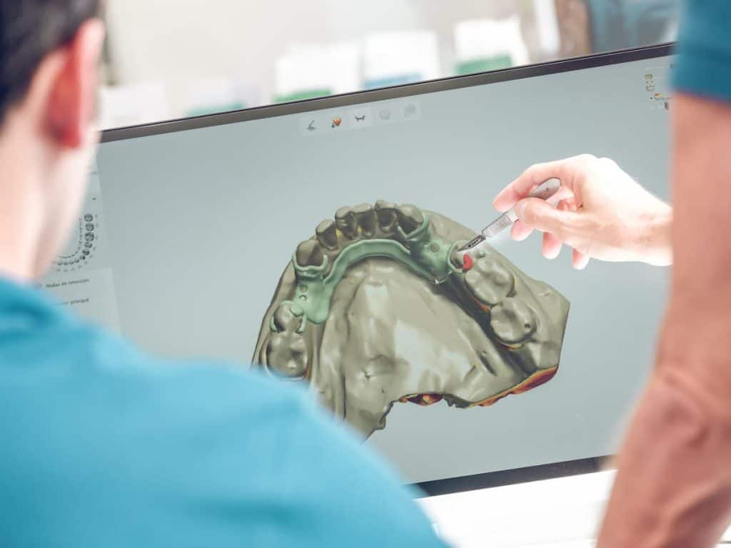
Some CBCT systems allow the dentist to create a virtual model of your mouth and simulate potential treatments. This feature is especially useful for implant planning, orthodontics, and other complex procedures. Patients can see a visual preview of what their outcome might look like before the procedure begins.
Applications of 3D CBCT
Dental Implants
CBCT is often used for pre-implant assessments. It helps determine bone quantity, quality, and location, and it guides implant placement, ensuring the best results and minimizing complications.Endodontics (Root Canals)
Dentists use CBCT to visualize the internal structures of a tooth when performing complex root canal treatments, especially if there is concern about extra or curved canals, fractures, or infection around the roots.Orthodontics
Orthodontists use CBCT to help plan tooth movement and jaw alignment for patients undergoing braces or clear aligner therapy. It also allows for a comprehensive view of the roots, bone structure, and the temporomandibular joint (TMJ).Oral Surgery
CBCT provides invaluable data for oral surgeons planning procedures like wisdom teeth extraction, jaw surgery, and tumor removal. It helps them assess bone structure, nerve proximity, and potential complications.TMJ and Airway Analysis
CBCT can be used to evaluate the TMJ (temporomandibular joint) and airway for patients with jaw pain or breathing disorders. It’s particularly helpful for diagnosing sleep apnea or other airway-related concerns.Cavity Detection and Tumor Screening
CBCT is used to detect hidden cavities, root fractures, or any suspicious growths or tumors that might not be visible on traditional X-rays.Safety of CBCT Imaging
While CBCT scans do involve exposure to X-rays, they use significantly lower doses compared to traditional CT scans. Modern CBCT machines are designed with safety in mind, and the amount of radiation is minimized as much as possible. As always, pregnant patients or those who might be pregnant should inform their dentist to assess the necessity of the procedure.